EN


Description:
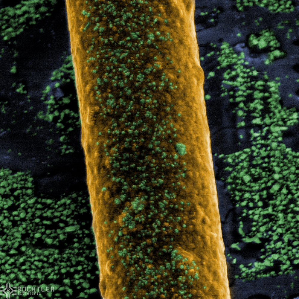
Scanning Electron Microscope Imiging allowes one to view the surface topology of samples at resolutions up to 2nm (currently highest resolution avalible at Bochtler Scientific). This allows the client to study surfaces in detail not avalible with the likes of the Light Microscope.
Additionally to just surface topology, elemental analysis of samples at high spacial resolution can be performed by means of Electron Beam Induced X-Ray Flourescence spectroscopy. This allowes the client to determine the chemical composition of the sample, and map the elements presence on the sample surface
Sample Preperation:
In order for objects to be viewed within the High Vacuum enviroment of the Scanning Electron Microscope, samples need to be specially prepared prior to imiging. Bochtler Scientific offers this preperation of all types of objects, both Biological and Non-Biological. Usually the sample to be viewed needs to be reduced in size to less then 1" in diameter and 1/2" in hight in order to be viewed within the microscope. This usually involves irreversable damage to the parent part due to having a small enough sub section extracted from it.
This section is then further proscessed, among which are drying and coating with a conductive layer. This layer can be made to be removable after imiging is completed, though this WILL reduce the maximum obtainable resolution. Normally samples, once prepared for imiging within the Electron Microscope can not be returned to their state prior to sample preperation.
Samples can be stored on site, or returned to the client, including the parrent part, if applicable, and the prepared SEM sample mounted on an appropriate sample holder. Survival of the prepared sample during shipping can not be garanteed, therefor it is reccomended that samples once prepared, be stored on site within the sample archive of Bochtler Scientific, should future analysis be needed.
Capabilities:
Our current Scanning Electron Microscopes are currently capable of the fallowing imiging methodes and sample sizes
| Point Resolution: | 2 nm |
|
Detectors: |
SED, BSED, EBIC, EDX |
| Maximum Sample size: | 1" x 1" x 1" |
Description:
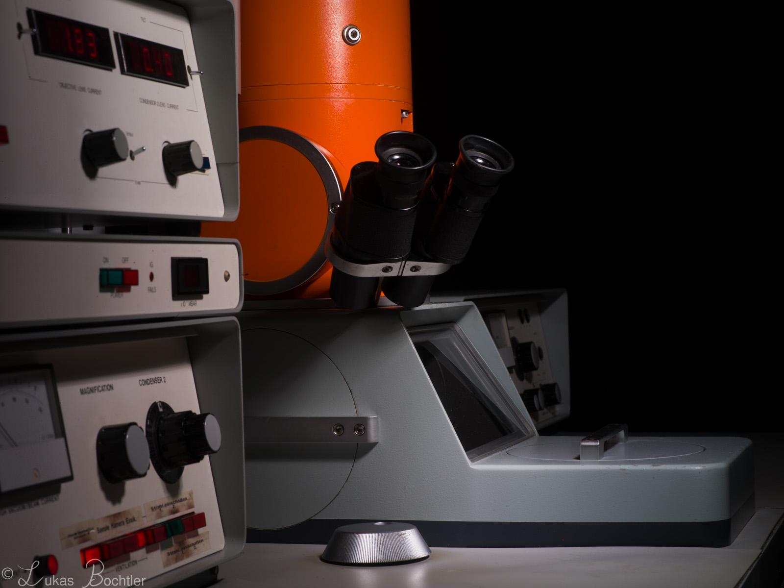
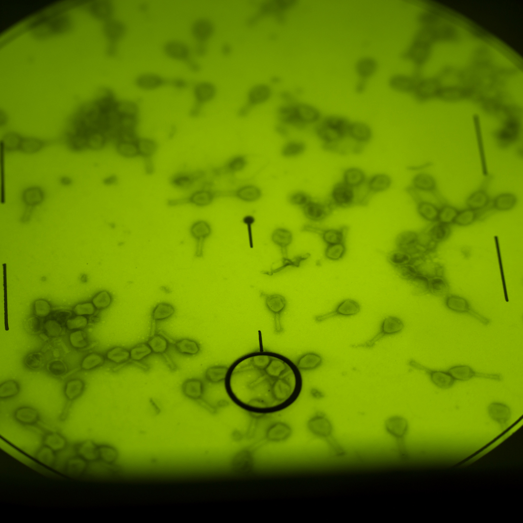
Transmission Electron Microscopy allows the observation and study of very thin layers of both Inorganic, Organic and Biological materials. Examples of structures visible within images created by the Transmission Electron microscope are, among others, Cell Organells, Viruses, Crystal Latices and Crystal Latice Boundrys. Additionally, elemental anylsis and dynamic phenomenon can be observed within the Treansmission Electron Microscope.
Sample Preperation:
Samples which are to be observed within the Transmission Electron microscope must be cut into very thin <70nm sheets no more then 2mm in diameter. This is acomplished by various means, dpeneding on if the subject is of biological, organic or inorganic origin, as well as what exactly is to be observed. Typcially sample preperation involves the proscessing of the subject into a state where it can be stabilized in a Expoy like resin. Additioanlly, depending on the sample, heavy metal salts my be added prior to embedding within the resin. The resulting resin blocks are then cut by means of a glass knife within a machine called an Ultramicrotome, which, by mechanicaly means, creates the <70nm thin sample sheets. Alternatively, ion Etching and or Ion Milling my be used to section a thin enough sample from the object under study. Usually the resulting samlpes are further proscessed in heavy metal salts, such as Uranium Acetate, Lead Citrate, Osmium Tetroxide, Phosphotungstik acid and the like. Due to the high toxicity of the resulting TEM sample, it is reccomended to have it remain within the Bochtler Scientific Archive after imiging. Samples can be shipped to the Client upon special request, though doing so is not reccomended.
Capabilities:
Our current Transmission Electron Microscopes are currently capable of the fallowing imiging methodes and sample sizes
| Point Resolution: | 5 Å |
|
Camera: |
4 MP |


Description:
Light Microscopes offer many ways of observing the microscopic world around us. Their resolution is rather limmited in comparison to the electron microscope, however they have the advantage of much simpler sample preperation and no requirement for samples to be water free.
Samples can be observed in basic manners, either by transmitting light through a semi transperent object, or by reflecting light off the surface of an object. The light path can be altered by many means to atain many different forms of information from the sample.
Additionally, with some samples and preperation methodes, the observation of flourecent behavior can be observed. This can be further combined with the Electron Microscope by means of Electron Induced Flourecence, such as is useful in minerology.
Due to the very shallow plain of focus within the light microscope, compared to that of the scanning electron microscope, taking numerous pictures of the sample under observation, each having this plain of focus shifted up or down by a certain amount relative to the last frame, a picture having increased depth of field akin to those from the Scanning Electron microscope, can be digitally reconstructed.
Sample Preperation:
Depending on the observation methode chosen, and the goal of the observation, different preperation methodes are performed.
In transmitted light microscopy, if the sample permits this, such is the case with biological samples, a small section of the sample is extracted from its bulk. This is then further chemically and or physically treated to allow it to be cut into thin section ranging in thickness from some tens of um to as thin as 500nm. These are then further treated with chemicals (stains) to increase contrast and or differentiate certain parts of the structure from one another. Alternatively the samples my be viewed by means of interference or phase contrast. These methodes have the advantage that even living subjects my be viewed.
For Flourecence microscopy the samples are prepared as above, but treated with different stains and or antibodies to visualize proscessed within cells. Some samples exibit the ability to flourece on their own, such is the case with phosphors, or the chloroplasts in plant leafs. This flourecence can also be pumped by means of an electron beam, this is used in among other fields, mineralogy.
Sample preperation for Reflected light microscopy is somewhat simpler, allowing even the observation of surface details on the unaltered bulk sample. However, for conviniance it is usually a good idea to extract a smaller volume from the bulk sample for observation.
In the case of metals, and metal structures, it is sometimes necessery to flatten and polish the surface to be observed. this surface is further treated with an etchant to reveal, for example, the grain boundrys within bulk metals.
A somewhat rare but useful imaging methode for reflected light microscopy is the Magnetooptic Kerr microscope, which is capable of visualizing the magnetic field present at the surface of the sample. This technique can be used to, for instance, retrieve information on damaged magnetic media which is damaged to such an extent, that it is no longer readable by the playback device, or is to delicate and or valuable to risk playback with normal magnetic read heads.
Capabilities:
At Bochtler Scientific, we offer many light microscopy techniques, these include, but are not limmited to:
- Transmitted Light Bright Field Microscopy
- Transmitted Light Dark Field Microscopy
- Transmitted Light Phase Contrast Microscopy
- Epi Flourecence Microscopy
- Reflected Light Bright Field Microscopy
- Reflected Light Dark Field Microscopy
- Reflected Light Interference Microscopy (DIC)
- Transmitted Light Bright Field Microscopy
- Transmitted Light Dark Field Microscopy
- Transmitted Light Phase Contrast Microscopy
- Epi Flourecence Microscopy
- Reflected Light Bright Field Microscopy
- Reflected Light Dark Field Microscopy
- Reflected Light Interference Microscopy (DIC)
Services
Copyright © 2021 - all rights reserved - Bochtler Scientific